6 Times Giant Eye Model Manufacturer,Supplier and Exporter in India

Product Code : EDS-HPM-12822
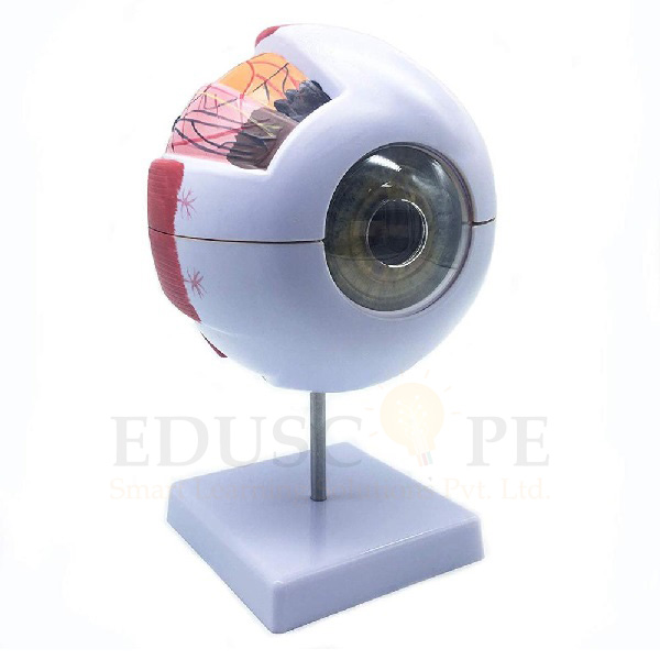
Eduscope India is a leading 6 Times Giant Eye Model Manufacturer,and suppliers in India, 6 Times Giant Eye Model Manufacturer,and suppliers in South Africa, 6 Times Giant Eye Model Algeria (Algiers), 6 Times Giant Eye Model Angola (Luanda), 6 Times Giant Eye Model Argentina (Buenos Aires), 6 Times Giant Eye Model Armenia (Yerevan), 6 Times Giant Eye Model Australia (Canberra), 6 Times Giant Eye Model Austria (Vienna), Bahrain (Manama), Bangladesh (Dhaka), Bhutan (Thimphu), Bolivia (Sucre), Botswana (Gaborone), Brazil (Brasília), Brunei (Bandar Seri Begawan), Montenegro (Podgorica), Morocco (Rabat), Mozambique (Maputo), Myanmar (Naypyidaw), Namibia (Windhoek), Nepal (Kathmandu), New Zealand (Wellington), Nigeria (Abuja), Oman (Muscat), Palestine (Ramallah), Panama (Panama City), Papua New Guinea (Port Moresby), Paraguay (Asunción), Peru (Lima), Philippines (Manila)¸ Portugal (Lisbon), Qatar (Doha), Rwanda (Kigali), Saudi Arabia (Riyadh), Senegal (Dakar), Serbia (Belgrade), Sierra Leone (Freetown), Slovakia (Bratislava), South Africa (Cape Town) (Pretoria) (Bloemfontein), South Sudan (Juba), Spain (Madrid), Sri Lanka (Sri Jayawardenepura Kotte) (Colombo), Sudan (Khartoum), Syria (Damascus), Tanzania (Dodoma), Thailand (Bangkok), Togo (Lomé), Tonga (Nuku'alofa), Trinidad and Tobago (Port of Spain), Tunisia (Tunis), Turkey (Ankara), Turkmenistan (Ashgabat), Uganda (Kampala), United Arab Emirates (Abu Dhabi), United Kingdom (London), United States (Washington, D.C.).

Description :
Enlarged. 
About 6 times of life size. Comes with a horizon section of the eyeball wall. 
Sclerotic membrane, choroids membrane, lens and vitreous humor are removable. 
The blood supply and the origins of the muscles control the eyeball movement are also shown. 
On stand with base. 6 parts.

Features :
6X Enlarged Human Eye Model on base for use in patient education or anatomical study. the model display the anatomical structure of human eyeball, such as the three layers of outer membrane (outer membrane, media and intima) of eyeball wall and the main refractive bodies, lens and vitreous body filled inside. 
The model includes a labeled diagram and is about 9.8x6.2 x 6.2 inches (H x W x D , where H is height, the vertical distance from the lowest to highest point; W is width, the horizontal distance from left to right; D is depth, the horizontal distance from front to back). 
Anatomical models are typically used as educational aids in medical and scientific classrooms and office settings.
The different parts of the eyeball model are detachable to show the following structures. 
1. Tunica external : showing cornea and sclera with attachments of ocular muscles and optic nerve. 
2. Tunica media : showing the iris, the ciliary body and the chorioid. 
3. Tunica internal is retina. 
4. Refraction media : showing the lens and the vitreous body. 6 times enlarged. On stand.


Office Address

Works: EDUSCOPE SMART LEARNING SOLUTIONS PRIVATE LIMITED,500, 5th Floor, ITL Twin Tower,Netaji Subhash Place, Pitampura,
Delhi – 110034, India,

[email protected]

Follow Us